A gutty experience
Anatomy students dissect rats to gain hands-on knowledge
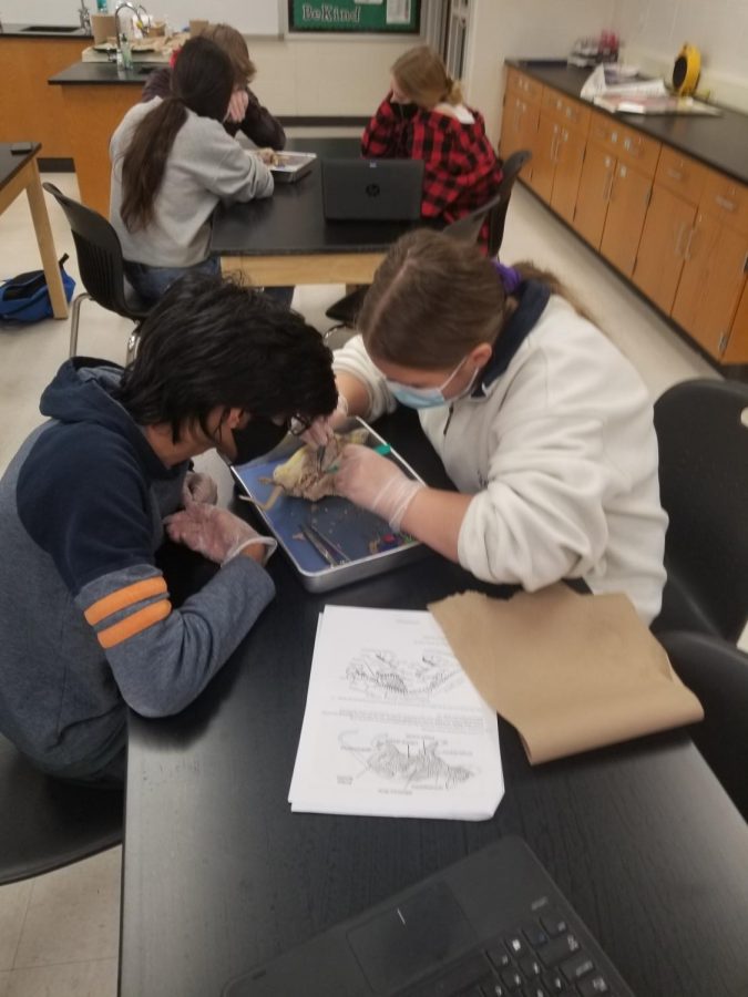
Students cut into the rat and followed the directions on their lab sheet. “We had to follow a guide that was step by step to dissect the rat,” junior Gina Olson said. “We could also ask the teacher for help if we needed it.”
December 18, 2020
The lab table is filled with tools of all different shapes and sizes. Students get ready to cut into the skin and discover the internal organs and muscles of the rat. Students spent Dec 10 and Dec 11 getting a hands-on experience as to what the body looks like internally.
Both McIver and the students thought that the dissection was a valuable experience, and the students learned many lessons from the dissection. They had been learning about the different organs and structures of a human all semester, and the rat dissection was a hands on experience that summed the semester up. They used rats because the rat has similar structures and functions as the human.
“We have many diagrams and models we use to study organs and organ systems, but seeing the real thing is such a different and more meaningful experience,” Anatomy teacher Amy McIver said. “Many students are surprised to see what the real organs look like. Also, the class is structured so that we study each individual body system and although we discuss how each system is related to other systems, the rat is a visual of all the systems put together. The relationships can be seen much more clearly in a real specimen.”
For students, they enjoyed analyzing the organs and comparing them to the organs of humans. Since the rat is a vertebrate, it has a backbone, just like humans do. Students loved getting to experience an animal firsthand.
“My favorite part was looking at all the organs and comparing them with our organs,” junior Gina Olson said. “I like learning about animals hand on. It really helps me visualize the different parts, and I can better understand it.”
The process of doing the dissection was very complex, and students had to follow very specific instructions as to how to cut open the rat properly. If the instructions aren’t followed correctly, the organs could potentially be damaged.
“They begin by identifying different regions of the rat using the anatomical terminology they have learned in class,” McIver said. “Next, they will remove the skin and identify specific muscles–many of which humans also have. Next, the students will begin looking at structures in the head and neck, like the salivary glands and trachea. Now it is time to cut into the abdomen and thoracic cavity and identify many more of the internal organs and observe their relationship to each other and discuss how that affects their functions. Finally, the students will explore the excretory and reproductive systems.”
Students determined the sex of their rat when they analyzed the reproductive system. Groups with the same sex rat will pair up to identify the rat’s reproductive organs and they will analyze how rats have children. If they have a female rat, they will then look at the differences in rat reproductive systems and human reproductive systems. Some students had a pregnant rat, and they looked at how the baby rat develops compared to how a baby develops in a human.
This was a valuable experience for students, especially ones that want to go into medical school. Having this class and the hands-on experience on their resume helps with getting into medical school.
“The body is incredibly complex, and when we are told that we’re doing something really well, it’s motivating because it validates skills applicable to premed and medical school,” senior Genevieve Graner said. “Experience in these particular assets really matters because premed and med school are so competitive, and we need all the edge we can get.”
Overall, the students had a lot of fun with this project, and it was a really good experience. Even if medical school isn’t in their future, they can still learn a lot from this experience.


